Dog Red Eye Emergency Triage: Squinting, Cloudiness, Trauma, and Safe Transport
A conservative veterinary triage guide for dog eye redness, squinting, cloudiness, trauma, exposure, and safe emergency transport without home diagnosis or treatment.
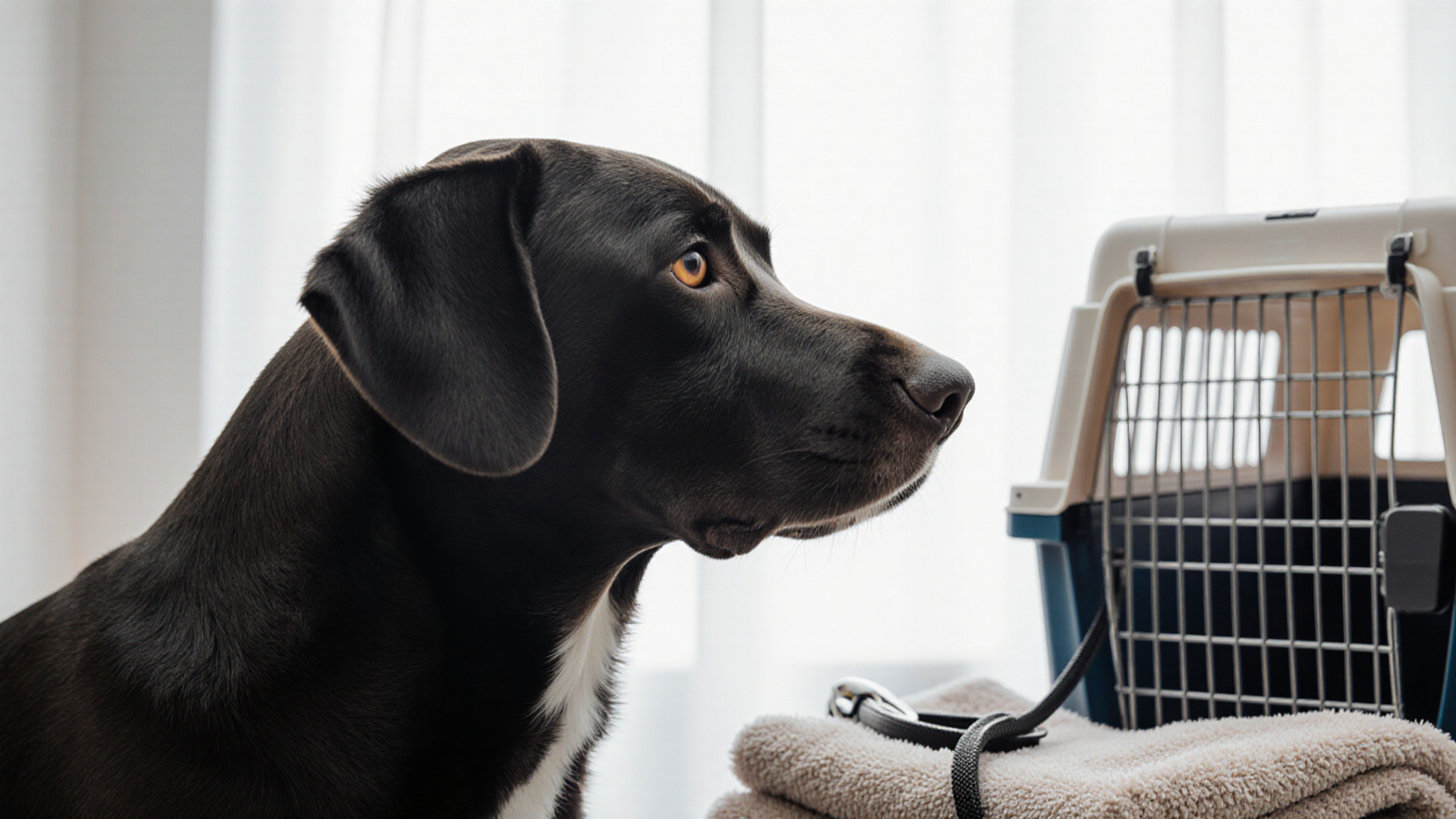
A dog’s red eye can reflect irritation, but the same outward color can accompany painful, vision-threatening disease. At home, you cannot reliably distinguish a superficial problem from glaucoma, a corneal ulcer, bleeding, a foreign body, lens trouble, or deeper inflammation. The Merck Veterinary Manual overview of ophthalmic emergencies emphasizes rapid recognition and stabilization because delay can affect comfort, vision, and sometimes the eye itself.
This guide is intentionally conservative. It supports triage, observation, calling, and transport; it does not diagnose the cause or teach eye treatment. Do not wait for a photograph to look dramatic. Pain behavior, abrupt change, injury, and loss of normal vision can matter more than the amount of visible redness.
Start with immediate danger, not a diagnosis
Move the dog away from traffic, stairs, other animals, broken glass, chemicals, smoke, and bright light. Keep your face away from the dog’s mouth. Even a gentle dog may bite when frightened or in severe eye pain. Do not force the eyelids open, press on the globe, pull at the third eyelid, or compare pressure by touching both eyes.
Call an emergency veterinary service now if you see any of these signs:
- an eye that suddenly looks enlarged, bulging, displaced, collapsed, or misshapen;
- severe or continuous squinting, crying, frantic pawing, or inability to open the eye;
- new blue, white, gray, or hazy cloudiness over the normally clear surface;
- sudden bumping into objects, missing steps, startling on one side, or other possible vision loss;
- a cut, puncture, bite, cat scratch, high-speed debris strike, blunt impact, or embedded object;
- blood inside or around the eye, a torn eyelid, or visible tissue protruding;
- chemical splash, corrosive powder, smoke exposure, or an unknown substance in the eye;
- unequal pupils with acute pain, collapse, neurologic change, or recent head trauma;
- rapid worsening over minutes or hours, especially with vomiting, profound restlessness, or lethargy.
The Merck glaucoma guide describes glaucoma as increased pressure inside the eye and notes signs such as pain, redness, cloudiness, and enlargement. Those signs overlap other conditions, so the safe household decision is urgent examination, not pressure guessing.
Redness alone is not a severity meter
“Red eye” may mean enlarged vessels on the white of the eye, inflamed conjunctiva, blood, red tissue, or reflection from a photograph. Note where the color appears without manipulating the eye. Also record whether discharge is watery, mucus-like, or bloody; whether one or both eyes are affected; and whether the change was sudden.
Squinting is a pain signal until a veterinarian establishes otherwise. A dog that holds one eye partly closed, repeatedly blinks, avoids light, rubs the face, or resists head touch should not be treated as having a cosmetic problem. Corneal disorders can be painful and require an examination that may include dye and magnification; the Merck overview of corneal disorders explains that ulceration and other corneal disease have multiple causes and presentations.
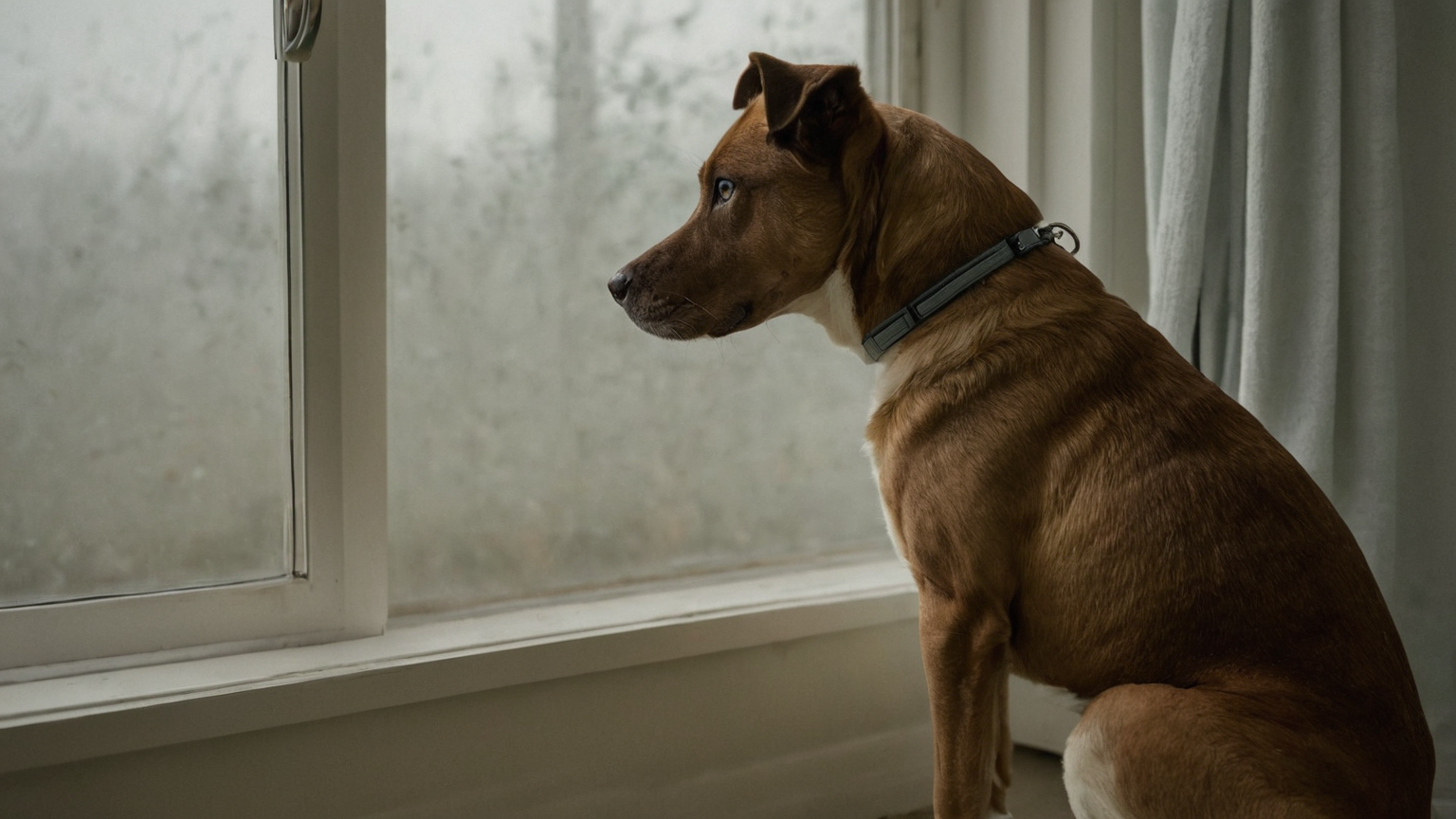
Cloudiness is particularly difficult to locate at home. It might be on the cornea, within the front chamber, in the lens, or deeper. A gradual age-related lens change is not a safe explanation for a newly painful, red, or asymmetric eye. UC Davis lists glaucoma, injuries, infections, cataracts, dry eye, and other conditions among problems evaluated by its veterinary ophthalmology service, using specialized examinations rather than appearance alone.
Decision table: call, leave, or monitor under direction
| Situation | Action now | Do not do |
|---|---|---|
| Penetration, embedded object, globe displacement, major bleeding, or tissue protrusion | Call emergency hospital and transport immediately | remove the object, press, patch tightly, or manipulate tissue |
| Chemical or corrosive exposure | Move away from exposure, call a veterinarian or poison service immediately, and follow product-specific professional instructions | improvise neutralizers or delay while searching social media |
| Sudden cloudiness, severe squinting, enlarged eye, sudden vision concern, or major trauma | Emergency assessment now | use leftover drops or wait overnight to “see if it clears” |
| Torn or bleeding eyelid without obvious globe damage | Urgent same-day/emergency assessment | trim tissue, apply glue, or put ointment into the eye |
| Mild redness with no pain, trauma, cloudiness, discharge, or vision change | Call the regular veterinarian for prompt triage | assume allergy or begin human medication |
| Already examined and stable under a written veterinary plan | Follow that exact plan and its recheck threshold | reuse the plan for the other eye or a future unrelated episode |
This table does not downgrade a case. A clinic may recommend earlier transport based on breed, age, prior eye disease, only-eye status, surgery history, medication, or the event description. VCA’s common canine emergency overview similarly places first aid after immediate safety and veterinary contact rather than presenting it as definitive treatment.
What not to put in the eye
Do not use human “get the red out” drops, allergy drops, antibiotic drops, contact-lens solution, essential oils, herbal washes, ointment, peroxide, or leftover medication. Do not use another animal’s prescription. Most importantly, do not use a steroid-containing eye product unless the veterinarian has examined this episode and directed it; some corneal problems can worsen with inappropriate medication.
Plain water or saline is not a universal home prescription either. Chemical exposures differ, and a puncture or unstable globe should not be flushed or pressed. Call while moving the dog away from ongoing exposure. The ASPCA’s Animal Poison Control resource provides 24-hour toxicology consultation in the United States, but a poison call does not replace veterinary examination of an injured eye.
Do not give human pain relievers. Do not feed a large meal while emergency anesthesia or sedation may be possible; ask the receiving hospital what to do about food, water, and routine medications. Never delay departure to complete a home remedy.
Prevent rubbing without creating pressure
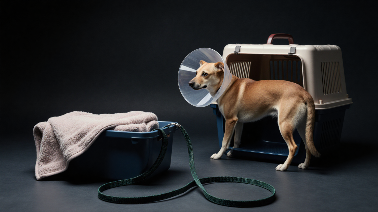
Rubbing can convert a painful surface problem into greater injury. If the dog already has a correctly sized rigid Elizabethan collar and it can be placed without touching the eye or causing a struggle, use it. The rim should extend beyond the nose. An inflatable collar may not prevent a paw from reaching the face, and a tight makeshift cone can obstruct breathing or press the neck.
Do not bandage the eye. Do not place gauze, cotton, or a cold pack directly over it. If an object protrudes, do not shorten or stabilize it with pressure; minimize movement and ask the emergency team for real-time instructions. The general AVMA first-aid guidance reinforces that first aid is not a substitute for veterinary care and that injured animals may react unpredictably.
Make a useful two-minute observation record
Record facts before memory becomes blurry, but do not delay transport:
- time the eye was last known normal and time the change was first noticed;
- which eye is affected and whether the other eye looks different;
- squinting, rubbing, light avoidance, discharge, cloudiness, bleeding, pupil difference, or vision behavior;
- exact mechanism: branch, cat claw, dog fight, vehicle debris, fall, shampoo, cleaner, aerosol, or unknown;
- product name, concentration, label photo, and exposure time when a substance is involved;
- all current medications, eye history, prior surgery, known glaucoma, and whether this is the dog’s only seeing eye;
- actions already taken, including anything placed in or around the eye.
Take one distant photograph only if safe. Turn off flash. Do not restrain the head, force an eyelid open, or repeat images while the dog struggles. A picture cannot measure eye pressure, stain a corneal defect, assess internal inflammation, or establish vision.
Call ahead and describe the highest-risk fact first
Say, “A branch struck the right eye and an object may still be present,” or “The eye became cloudy and the dog cannot open it,” before giving background. Ask which entrance to use and whether staff want you to remain in the vehicle. Confirm estimated arrival time and request handling instructions for a large, fearful, or painful dog.
Use the pet emergency go-bag guide to keep records and a leash ready before emergencies, but leave optional packing behind when vision may be threatened. If another adult must care for children or pets, the emergency foster-care handoff plan can shorten departure without mixing clinical instructions.
Safe transport checklist
- Call the receiving clinic and begin travel when directed.
- Keep the dog in a dim, quiet area; avoid direct air vents and open windows.
- Use a secure carrier for a small dog or a harness and nonslip surface for a larger dog.
- Keep paws away from the face with an appropriate cone when safely available.
- Keep hands away from the mouth and injured eye.
- Have one adult drive and another observe if possible.
- Bring medication names, exposure packaging, the timeline, and relevant records.
- Do not let the dog ride with its head out the window.
- Do not attempt drops, food, or treatment while the vehicle is moving.
- If the dog collapses, develops breathing trouble, or deteriorates, update the hospital immediately.
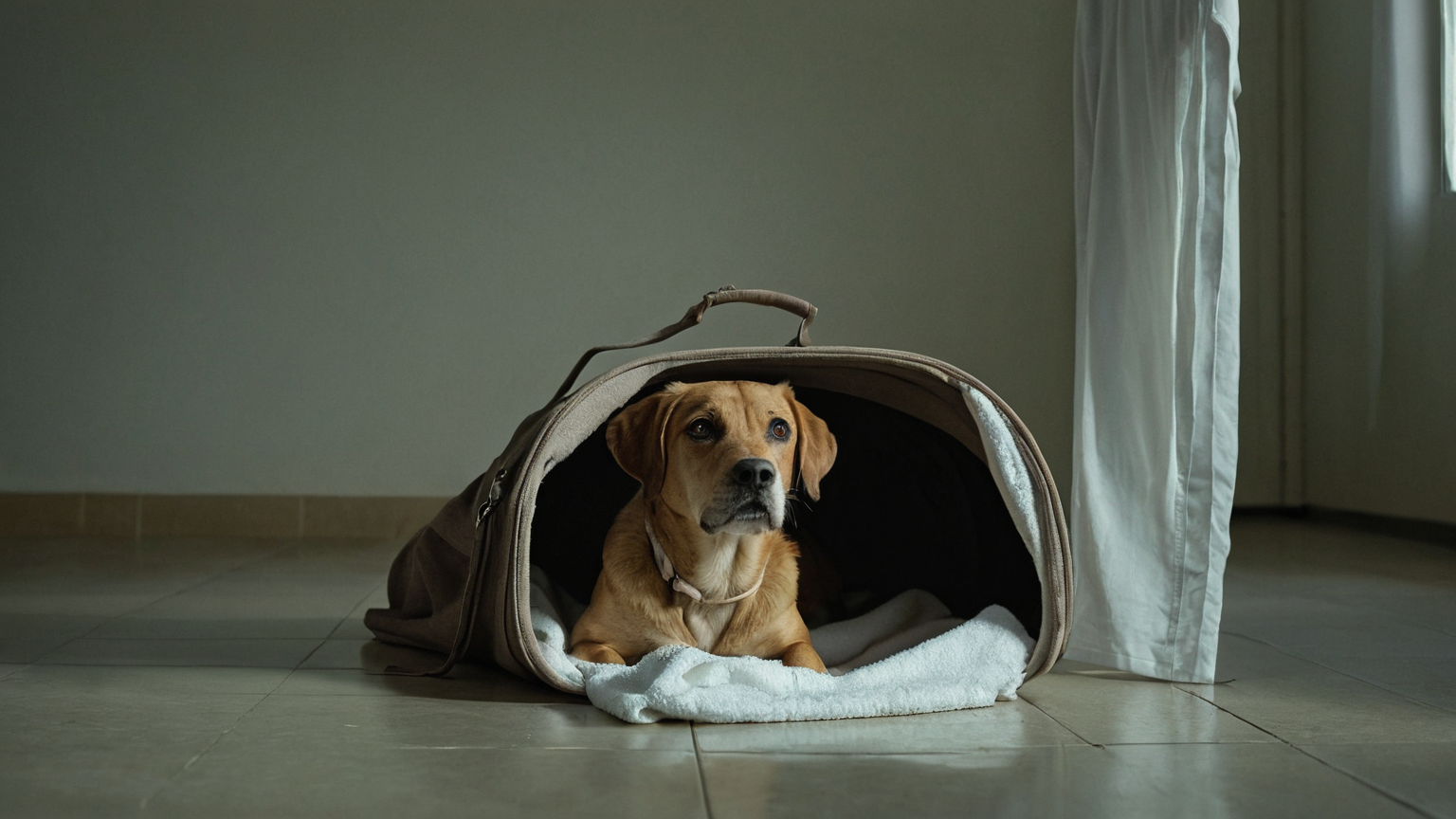
The broader loading principles in the dog GDV emergency transport plan can help with route and vehicle preparation, but eye emergencies have different handling boundaries: never press the head or eye against a blanket and never treat one emergency article as a diagnostic checklist for another.
Special exposure and heat boundaries
After a cleaner, pesticide, medication, plant sap, or unknown liquid reaches the face, protect yourself first. Ventilate the area, avoid bare-hand contact, and bring the intact label or a photograph. Do not mix chemicals to “neutralize” them. A veterinarian or poison specialist may give immediate decontamination directions based on the substance; follow those directions exactly while arranging examination.
Heat exposure can produce red mucous membranes, weakness, and neurologic signs that coexist with eye irritation. If overheating is possible, use the emergency thresholds in the dog and cat heatstroke first-aid guide while contacting a veterinarian. Do not focus on the eye and miss collapse, abnormal breathing, or dangerous body heating.
What the veterinary team may need to determine
The clinic may assess vision responses, eyelids, tear production, corneal integrity, eye pressure, pupils, the front and back of the eye, and injuries beyond the globe. Tests and sequence depend on suspected trauma and stability. A general veterinarian may treat the emergency, consult, or refer. The American College of Veterinary Ophthalmologists provides an ophthalmologist search, but referral logistics should not delay the nearest appropriate emergency assessment.
The absence of one test does not mean the visit was incomplete; penetrating injury or severe pain can change what is safe initially. Conversely, a normal-looking photograph cannot substitute for an examination. Ask for written instructions: medication name and eye, exact schedule, cone use, activity limits, recheck time, and signs requiring immediate return.
Limitations and safety boundary
No home checklist can identify the cause, depth, pressure, infection status, or prognosis of a red eye. Symptoms can overlap, dogs may hide vision loss, and both eyes may require examination even when one appears normal. Travel time and clinic availability do not make watchful waiting safer. If your veterinarian gives a more cautious instruction, follow it.
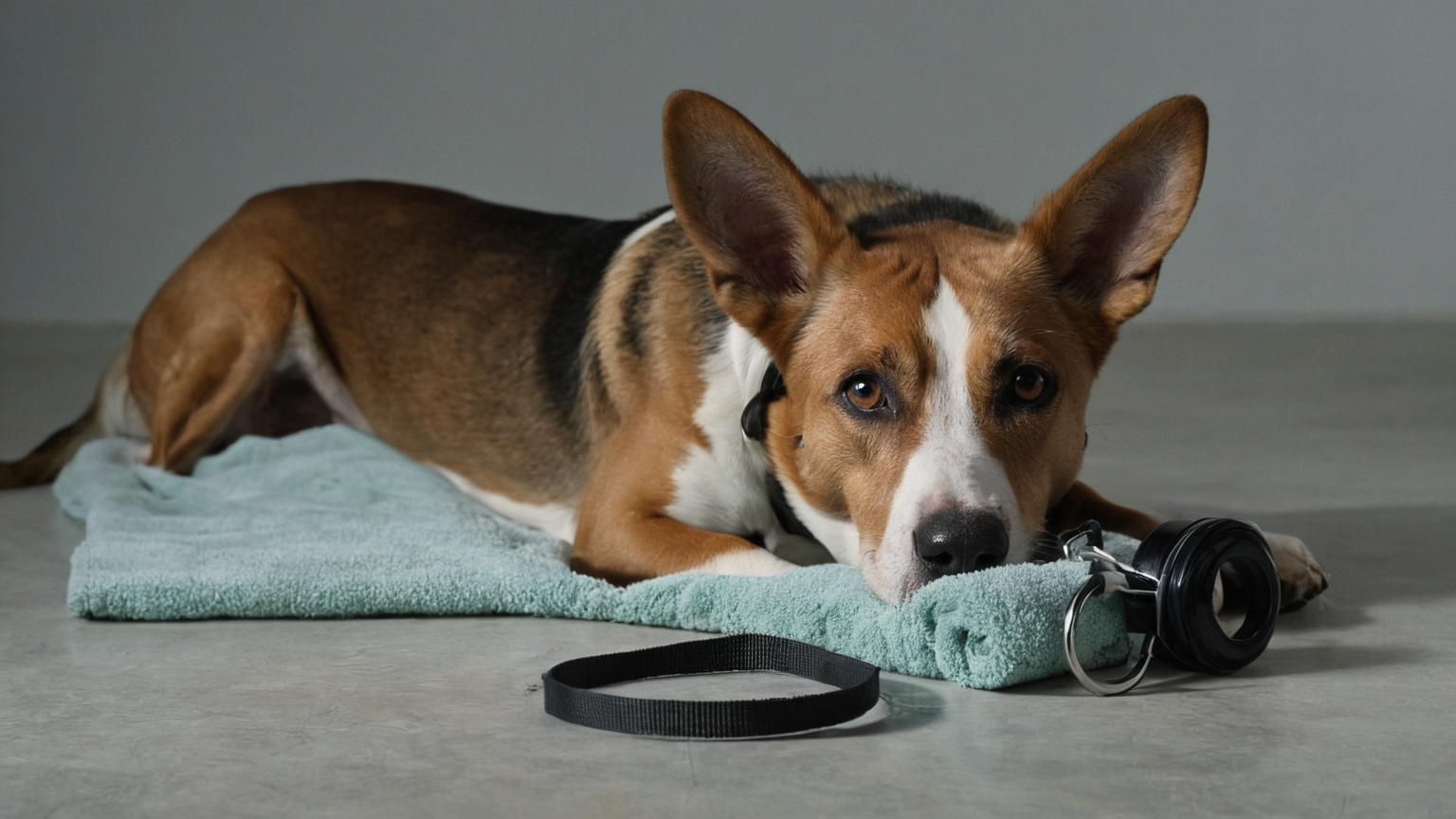
Final leave-now checklist
- severe squinting or obvious pain;
- sudden cloudiness, enlargement, shape change, or vision concern;
- trauma, bite, scratch, puncture, foreign object, or bleeding;
- chemical exposure or unknown substance;
- tissue protrusion, torn eyelid, or displaced eye;
- rapid progression or systemic illness;
- inability to prevent rubbing safely.
When in doubt, call an open veterinary service and describe the change. The safest household contribution is early recognition, no unapproved eye products, protection from rubbing and pressure, and calm transport—not a home diagnosis.